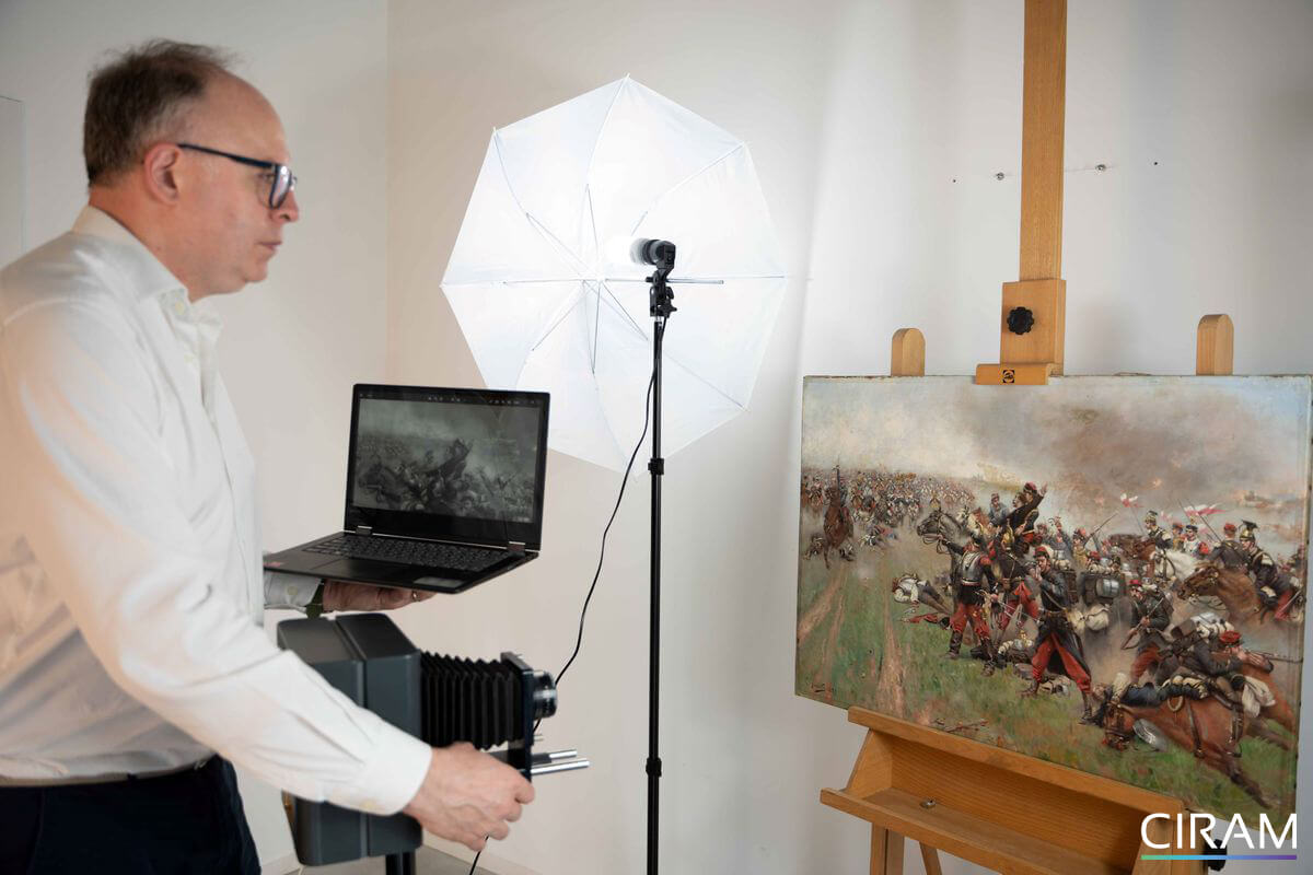


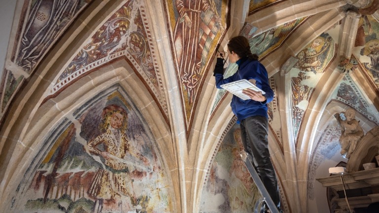
Scientific analysis of a painting makes it possible to examine the materials it is composed of in order to shed light on its history, state of preservation, and authenticity. Today, it is an invaluable tool for restorers, curators, collectors, experts, and professionals in the art market.
At CIRAM, this approach is based on complementary methods: dating the supports, chemical analysis of pigments, binders, and varnishes, as well as various scientific imaging techniques. Cross-referencing these results provides a better understanding of a work’s creation, helps identify any restorations or inconsistencies, and provides objective evidence for the purposes of the expert assessment.
In summary:
- Scientific analysis of a painting combines dating, chemical analysis, and imaging techniques to study the materials that make up the artwork.
- Scientific imaging (visible light, ultraviolet, infrared, and X-rays) reveals the different layers of a painting, as well as restorations and alterations that are invisible to the naked eye.
- These analyses make it possible to identify materials, assess the state of preservation, and provide useful information for authentication.
- The interpretation of the results is based on cross-referencing scientific data with historical and artistic knowledge.
The Main Methods of Scientific Analysis of Paintings
To study a painting, several complementary approaches are used. Some allow researchers to date the materials, while others help identify their composition or reveal elements invisible to the naked eye. These methods are chosen based on the nature of the artwork and the questions being addressed.
| Method: | What it allows us to study: |
| Dating of Media | Determining the Age of Organic Materials |
| Chemical Analyses | Identify the materials used and their historical consistency |
| Scientific imaging | View invisible layers, restorations, and modifications |
Dating the Media
Dating the supports makes it possible to estimate the age of the organic materials that make up a painting, such as linen or hemp canvas, wood, paper, or cardboard. Performed primarily using the carbon-14 method, this process provides a valuable chronological reference for verifying the consistency between the materials used and the presumed period of the work’s creation. However, this analysis does not directly date the paint or the creation of the work, but rather the moment when the organic material ceased to be alive—for example, when the flax was harvested or the tree used as a support was felled. The results should be interpreted in conjunction with other scientific and historical investigations.
The carbon-14 method is discussed in greater detail in our article on carbon-14 dating of paintings.
Chemical Analyses
Chemical analyses make it possible to identify the composition of the various materials that make up the paint layer. These analyses focus in particular on pigments, binders, varnishes, and mineral fillers. They help verify whether the materials are consistent with the work’s presumed period of creation, detect certain restorations, and provide additional information to support dating and scientific imaging.
The methods for identifying pigments, binders, and varnishes are detailed in our article on the chemical analysis of paintings.
Scientific imaging techniques
Scientific imaging provides valuable information about a painting. It allows for the examination of all layers of a painting—from the varnish to the support, including the preparatory drawing, the paint layers, retouches, overpainting, and any restorations. Natural light, ultraviolet, infrared, and X-ray imaging—discover the full range of imaging techniques used to analyze paintings.
These methods are completely non-destructive. Find out why this type of analysis is preferred for works of art.
Visible light imaging
We distinguish between three different types of visible light examination:
- Visible light photography: produced under specific conditions, these images correspond to classic photography;
- Grazing light photography: this type of photography reveals the state of conservation of the work's surface. Our scientists illuminate the work with a focused beam of light forming a 15° angle with its surface. This technique makes it possible to detect alterations to the paint layer (lifting, blistering, cracking, etc.), deformations of the support (poor tension of the canvas, cracks or joining of the boards) and alterations such as tears, scratches or dents. Grazing light also provides clues to the painter's techniques and style.
- Transmitted light photography: this is used to observe the reverse side of canvas or paper paintings. With this method, our scientists can detect holes and tears, as well as alterations in the paint layers.
Ultraviolet (UV) radiation
Ultraviolet (UV) radiation lies outside the visible spectrum, and is divided into near UV (380nm to 200nm) and extreme UV (200nm to 10nm).
UV imaging enables the surface layer of the painting to be explored. It uses the fluorescence properties of the varnish, which are more or less intense depending on its composition and alteration. Overpainting on varnish or restorations can be easily identified.
Important: in the absence of varnish, some natural pigments may emit a colored fluorescence, a fact to be taken into account to avoid misinterpretation.
Infrared (IR) radiation
Infrared radiation ranges from 780 nm to 5 mm, beyond the visible spectrum but on the opposite side of the ultraviolet spectrum. The CIRAM laboratories use what is known as “warm” light, which falls within the near-infrared range. This makes it possible to measure the absorption of infrared radiation by the materials that make up the paint layers.
Thanks to infrared reflectography, our scientists can reveal preparatory drawings, repentirs and repaints. This imaging method produces grayscale images corresponding to varying degrees of interaction with the initial radiation.
X-ray radiography
X-ray radiography captures an image of the internal structure of a painting. The X-ray beam is directed onto the painting, and the X-rays transmitted are observed as a function of the selective absorption of the material. This absorption is linked to two parameters:
- Through thickness
- The density of the materials making up the object.
With this examination, it is possible to identify the damage caused by the passage of time, as well as anthropic actions of all kinds from the creation of the work to the present day. It is also possible to identify repaints and repentirs thanks to contrasts in material density.
X-ray imaging is one of the most important imaging techniques. Discover how it reveals the hidden history of a painting.
The equipment used by CIRAM scientists
Visible-light imaging is performed using a 10-megapixel SAMSUNG® EX2F digital camera equipped with a 24–80 mm f/1.4–2.7 lens.
Infrared reflectography imaging is carried out between 900 nm and 1700 nm using an OSIRIS HD Infra Rouge Opus Instruments Ltd® 16-megapixel digital acquisition system. The equipment used for X-ray radiography is portable, comprising a mano-medical HF 1060 high-frequency X-ray generator, a FUJI D EVO II digital acquisition sensor with automatic triggering and Bluetooth transmission (FDR D-EVO, 35 x 43 cm), and a computer for generator control and image processing (OSIRIX software). The acceleration voltages used are 40 kV, 50 kV and 75 kV. Parameters were controlled using Fujifilm® software.
Using this state-of-the-art equipment, CIRAM’s laboratories date and analyze your paintings, sculptures, and ethnographic and archaeological artifacts.
Share this article

You may be asking yourself these questions about the scientific analysis of paintings?
No single method can, on its own, answer all the questions raised by a work of art. Dating provides information on the age of the materials, chemical analyses identify their composition, and imaging techniques reveal the various layers of paint as well as restorations invisible to the naked eye. Cross-referencing these results allows for a more reliable interpretation and helps minimize the risk of error during the authentication or study of a painting.
X-ray imaging allows us to examine the internal structure of a painting. It highlights differences in the density of materials and reveals elements invisible on the surface, such as the artist’s corrections, repainting, certain restorations, the construction of the support, and alterations resulting from the work’s aging. It is a valuable tool for understanding the painting’s technique and material history.
Restorations can be identified using several complementary methods. Ultraviolet light imaging often reveals repainting and certain varnishes, while infrared reflectography and X-ray imaging make it possible to observe interventions carried out beneath the visible surface. Chemical analyses of the materials complement these observations by identifying substances or pigments that are incompatible with the work’s presumed period of creation.
No. Scientific analysis provides objective data on a work’s materials, production techniques, state of preservation, and chronology. On the other hand, attributing a painting to an artist, studying its style, provenance, or historical context are matters for art historical expertise. These two approaches are complementary, and comparing them allows for the most reliable conclusions to be drawn.
Most scientific imaging techniques, such as photography, infrared reflectography, and X-ray radiography, are completely non-destructive. Certain analyses, however—including carbon-14 dating and some chemical analyses—require the removal of a very small sample of material. This is done with great care, from an inconspicuous area of the artwork, in order to preserve its integrity.
Related topics
Need more information or have a question?
Our team will get back to you as soon as possible.